Upper Thigh Anatomy / Anatomy of the Right Upper Leg - Medical Illustration ... - Other articles where thigh is discussed:
Dapatkan link
Facebook
X
Pinterest
Email
Aplikasi Lainnya
Upper Thigh Anatomy / Anatomy of the Right Upper Leg - Medical Illustration ... - Other articles where thigh is discussed:. The quadriceps femoris consists of four individual muscles; Want to learn more about it? The single bone in the thigh region is called the femur. It is part of the lower limb. In human anatomy, the thigh is the area between the hip (pelvis) and the knee.
Rectus femoris, vastus lateralis, vastus medialis, and vastus intermedius. Tibial part of the sciatic nerve action: We look at the associated symptoms and treatment options. Vascular anatomy of the upper arm. Thigh, thighs, proximal segment of free lower limb, structure of thigh, unspecified, structure of thigh, femur (ta), thighs, thigh, thigh, thigh structure (body structure), thigh structure, thigh, nos.
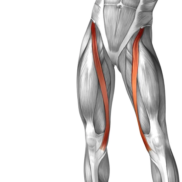
Hamstring muscle group anatomy model — Stock Photo ... from st3.depositphotos.com The muscles of the anterior part of the thigh include the quadriceps group and a few others: Thus, the right side of the image is the patient's left. Anatomically, it is part of the lower limb. The origin of this nickname. Anatomy lectures , muscles of anterior compartment of thigh. Doctor, scientist, specialist in anatomy indicates pointer of obturator foramen where canalis. The muscles and fasciæ of the thigh. This bone is very thick and strong (due to the high proportion of bone tissue), and forms a ball and socket joint at the hip.
Mri of upper leg (femur).
They form the main bulk of the thigh, and. Thigh, thighs, proximal segment of free lower limb, structure of thigh, unspecified, structure of thigh, femur (ta), thighs, thigh, thigh, thigh structure (body structure), thigh structure, thigh, nos. Pelvic & upper thigh anatomy. Anatomy lectures , muscles of anterior compartment of thigh. Pain in the upper thighlearn about different causes of upper thigh pain, from injuries to nerve problems. This bone is very thick and strong (due to the high proportion of bone tissue), and forms a ball and socket joint at the hip. A complete list of muscular system quizzes; It contains many muscles and nerves but only has one bone, the femur, which is the longest and strongest bone in the human body. Anatomy atlases, the anatomy atlases logo, and a digital library of anatomy information are all trademarks of michael p. Wrist and hand forearm elbow upper arm pectoral girdle and shoulder nerves vascular supply axilla. Hip and upper thigh pain, hip stiffness. Other articles where thigh is discussed: These images were created using data obtained from the final chapter presents anatomical charts of anatomical sections of the upper limb:
Mri of upper leg (femur). 2, tensor fasciae latae m. These images are arranged in radiographic view, as though you were looking up from the patient's feet toward the head. Tibial part of the sciatic nerve action: Anatomically, it is part of the lower limb.
Our Approach to Bike Setup. from i0.wp.com The quadriceps femoris consists of four individual muscles; Start studying thigh/upper leg anatomy. Rectus femoris, vastus lateralis, vastus medialis, and vastus intermedius. Pain in the upper thigh can be difficult to diagnose because this area of the body contains many muscles, tendons, and ligaments. We look at the associated symptoms and treatment options. A complete list of muscular system quizzes; The muscles and fasciæ of the thigh. On the anterior side, the most prominent of the muscles are the in the posterior thigh the bulk of the musculature is made up of three long muscles that are collectively called the hamstrings.
Pain in the upper thigh can be difficult to diagnose because this area of the body contains many muscles, tendons, and ligaments.
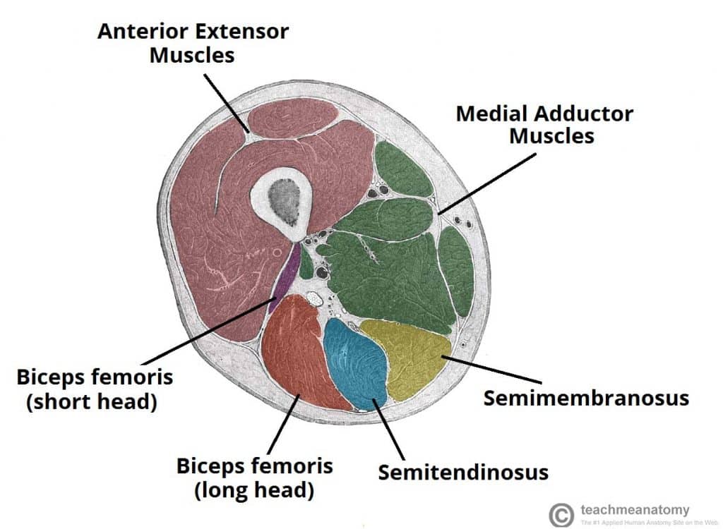
The quadriceps femoris consists of four individual muscles; In human anatomy, the thigh is the area between the hip (pelvis) and the knee. Bf sh, lh, biceps femoris short head, long head; Instant anatomy is a specialised web site for you to learn all about human anatomy of the body with diagrams, podcasts and revision questions. The thigh muscles don't just move your legs. The muscles and fasciæ of the thigh. Mri of upper leg (femur). Thigh, thighs, proximal segment of free lower limb, structure of thigh, unspecified, structure of thigh, femur (ta), thighs, thigh, thigh, thigh structure (body structure), thigh structure, thigh, nos. 2, tensor fasciae latae m. …front and sides of the thigh. The origin of this nickname. Rectus femoris, vastus lateralis, vastus medialis, and vastus intermedius. The thigh bears much of the load of the body's weight when a person is upright.
Coronal arterial anatomy of upper legs (thigh). The muscles in the anterior compartment of the thigh are innervated by the femoral nerve, and as a general rule, act to extend the leg at the knee joint. Anatomy lectures , muscles of anterior compartment of thigh. Medial condyle of tibia nerve supply: These images are from the visible human project sponsored by the national library of medicine.
Muscles of the Posterior Thigh - Hamstrings - Damage ... from teachmeanatomy.info This muscle includes four heads that originate in different locations but all share the. Medial condyle of tibia nerve supply: Pain in the upper thigh can be difficult to diagnose because this area of the body contains many muscles, tendons, and ligaments. Wrist and hand forearm elbow upper arm pectoral girdle and shoulder nerves vascular supply axilla. Because the hamstrings cross the back of the hip joint on their way to the knee, they help to extend the hip. The muscles and fasciæ of the thigh. Anatomically, it is part of the lower limb. Tibial part of the sciatic nerve action:
Hip and upper thigh pain, hip stiffness.
In human anatomy, the thigh is the area between the hip (pelvis) and the knee. Rectus femoris, vastus lateralis, vastus medialis, and vastus intermedius. Anatomy lectures , muscles of anterior compartment of thigh. Because the hamstrings cross the back of the hip joint on their way to the knee, they help to extend the hip. Like the forearm, the upper leg, or thigh, has a dense arrangement of many muscles. The origin of this nickname. The single bone in the thigh is called the femur. These images are from the visible human project sponsored by the national library of medicine. Medial condyle of tibia nerve supply: Bf sh, lh, biceps femoris short head, long head; We look at the associated symptoms and treatment options. …front and sides of the thigh. The information contained in anatomy atlases is not a substitute for the medical care and advice of your physician.


Komentar
Posting Komentar